Tattoo Rose - Sketches and Tattoo Rose
The meaning, history and value of the rose tattoo tattoo in the form of roses in western countries just like a lotus tattoo in ...
The tattoo of the goat, the photo of which are presented below, looks very eccentric and can be filled with a deep meaning. On the meaning of such a tattoo, as well as about what style it can be done, you will learn from this article.
Many may seem to make a tattoo with a goat is not worth it. However, tattoo art implies freedom of choice, and if you want to decorate your body in a similar way, then why not do it? True, first it is necessary to find out the symbolism of the goat tattoos: it can be different depending on the additional elements and even from the style of execution:
Tip! Before making such an ambiguous tattoo, it should be thicker to think about your decision. After all, the tattoo will remain with you for many years: it will not be easy to bring it out or overlapping it. In addition, the laser tattoo eliminates is a much more painful process than its application.

The tattoo of the goat, the sketches of which are presented in the article, can be executed in all existing styles. People, with respect relating to Satanism, can make such a tattoo in an engraving style. In this case, the image resembles an illustration to medieval treatises.
In the styles of Old-Skul and New School, the goat may look bright and optimistic. Add image to help ribbons with quotes, compasses, clocks and even flowers. Although such a combination may seem eccentric, the idea can be played quite interestingly.

It is possible to make a tattoo with a wild goat in a graphic style. The image looks as if it is based on the basis of the zoology textbook. Usually, the tattoo in the graphic style is complemented by bright color divorces that resemble watercolor "blots" and geometric patterns that make tattoo completed.
A realistic goat is a tattoo on which not everyone will decide. However, such work will surely attract a lot of attention to its owner. You can also choose Tresh-Polka style, involving expression, conciseness and style eclectics. Tresh polka allows you to beat complex philosophical ideas in graphic terms.

Tip! Decide with the style of the tattoo is not easy. Show the master workman who seem attractive to you, or even bring him aesthetic from your point of view illustration. This will help you decide on the choice of a suitable style for you.
Choose a place to apply a tattoo with the image of a goat with caution, because this motive can perceive very ambiguously. Bold nature, non-conventional, can choose open areas of the body, such as hands or feet. If you are afraid that the presence of a tattoo can affect your career and social position, better choose parts of the body that can be hidden under clothing. Then about the presence of your tattoo will know only close people. Special caution should be taken if you plan to make a tattoo with a goat, personifying the commitment of Satanism. After all, glances can change over time, and it will be very difficult to get rid of the tattoo.

Anatomy is a science that studies forms, structure, relationships and location of parts of the body, and physiology is a science that studies in the living organism processes (functions) and their patterns. The general data of these sciences will help you understand, for example, how to properly provide veterinary care to a sick animal.
The body of any animal is built of smallest living particles - cells. Certain cell groups, changing their shape and structure, are combined into separate accumulations that have adapted to performing certain functions. Such cell groups tend to have specific qualities and are called tabs. There are four types of fabrics in the body: epithelial, coupling, muscular and nervous.
Epithelial fabric It covers in the body all border formations, such as leather, mucous membranes and serous shells, output gying glands, gland internal and external secretion. It communicates with the body with an external environment, performs a coating, ferrous (secretory) and a suction function.
Connective tissue It is divided into feeding and reference. The supply, or trophic, fabric includes blood and lymph. The main purpose of the support tissue is to bind to a single integer component parts of the body and in the formation of the core of the body (for example, there are bone tissue, tendons and cartilage).
Muscle It is capable of reducing and relaxing under the influence of various stimuli. It is divided into a skeletal and heart muscles, which has cross-striped allocations, as well as smooth muscle tissue capable of involuntary contractions and occurring in the internal organs.
Nervous fabric It consists of neuron nerve cells with the property of the excitation and nervous excitation, and neuroglia cells that perform the support, trophic and protective functions.
Separate tissue groups are connected to each other and form organs. . Organ They call a part of the body having a certain external form constructed from several naturally combined tissues and performs any narrow specialist function. For example, the organ is called eye, kidney, language.
Separate organs performing together any one specific function form in the body systems,or Apparators. For example, bones, muscles, ligaments, tendons, joints form a movement device, or a musculoskeletal system.
The digestive, breathing, urinary and sexual organs, that is, the insides are located in three cavities: chest, abdominal and pelvic.
Breast cavity Located inside the chest, abdominal The front is limited to the diaphragm (breasting muscular barrier), and the rear goes into the pelvic cavity. It ends at the loan level. Pelvic cavity Form the bones of the pelvis, the sacral bone and the first tail vertebrae.
Most of the internal organs are located in the serous cavities that create conditions for the slip of the organs near each other. For example, the heart is located in a windowless serous cavity.
A prerequisite for the existence of any animal organism is metabolism - A continuously flowing process of decaying the components of the body, accompanied by the recovery process using the focus from the external environment. The metabolism and the conversion of energy in a living organism are inseparable from each other. Education and heat release depend primarily from metabolism. So, finely horned cattle - warm-blooded animals, that is, they have a relatively constant body temperature: in adult sheep - 38.5-40.0 ° C, a lamb - 38.5-40.5 ° C, in an adult goat - 38 , 5-40.5 ° C, and at the goat - 38.5-41.0 ° C. The body temperature depends on climatic and other factors, but most of all it changes under the influence of pathogenic microbes and viruses.
The body of sheep and goats, like other animals, is conventionally divided into four main departments (Fig. 3).
Fig. 3. Goats: Goats:1 - forehead; 2 - nose; 3 - nostrils; 4 - upper lip; 5 - Lower lip; 6 - chin; 7 - themes; 8 - heads; 9 - throat; 10,11 - neck; 12 - chest; 13 - withers; 14 - spin; 15 - loins; 16 - croup; 17 - hungry pits; 18 - chest; 19 - belly; 20 - groin; 21 - udder; 22 - the blade region; 23 - shoulder area; 24 - the area of \u200b\u200bthe elbow joint (elbow); 25 - forearm area; 26 - wrist area; 27 - Puta region; 28 - Went region; 29 - hoof; 30 - hip region; 31 - the area of \u200b\u200bthe knee joint (knee); 32 - the roof area; 33 - the area of \u200b\u200bthe jumping joint; 34 - area plus; 35 - tail.
Head. It distinguishes brain (skull) and facial (muzzle) parts. These include forehead, nose, ears, teeth.
Neck. Here they allocate the upper part (left region), the lower area of \u200b\u200bthe neck and the region of the jugular gutter (located above the trachea, where jugular veins are held).
Torchishche. It is represented by the withers (it is formed by the 5 first breast vertebrae and those with them at one level the upper edges of the blades), with a back, lower back region (breast), pyrical, crop, right and left iliac region, right and left steam, umbilical area, Udder region? or breast and prepitation, anal area, tail.
Limbs. Breast (front) limb is represented by shoulder, elbow, forearm, wrist, bowl; And the pelvis (rear) - the thigh, knee, shin, heel, plus.
The appearance of the animal, the physique and the features of the individual parts of the body, characteristic of the breed and the floor, are called exterior. The total exterior includes the main signs of the physique, the structure of individual parts of the body, as well as the most characteristic deviations and vices; The private exterior considers the features of the physique of individual rocks, their typical and nonypical signs. So, at the cattle of the dairy direction, the body is long, the backbone is thin, the head is small, a donkey rounded. The torso of small cattle meat referrals is compact, wide and deep, on relatively short legs.
The concept of " constitution»Combines all the properties of an animal organism: the features of its anatomical structure, physiological processes, and above all, the features of the highest nervous activity determining the reaction to the external environment. In Zootechny, 4 types of constitution are distinguished: rough (for example, meat breeds of sheep), gentle (for example, sheep with very thin wool), dense, or dry (for example, wool and cracked sheep breeds), loose, or raw (for example, meat sources sheep).
The type of higher nervous activity is closely related to the basic functions of the body - the metabolism, adaptability and a kind of reaction to the environment. All these reactions, in turn, are reflected in the exterior forms, which should be considered as an external reflection of the Constitution.
When determining the constitution of animals and the evaluation of the exterior establish a condition. Condition -this is a general view of the animal, external signs, fatness, musculature, skin, which helps determine whether the animal is great.
The movement apparatus is represented by skeleton, ligaments and muscles, which, unlike other systems, form the physique of small cattle, its exterior. To present its meaning, it is enough to know that the newborn on the device moves approximately 70-78% of the entire weight of the animal, and in adults - up to 60-68%. In phylogenesis, various departments are formed: a skeleton as a supporting structure, ligaments that provide bones and skeletal muscles that lead bone levers.
Bone - part of the skeleton, the organ, which includes various tissue elements. It consists of 6 components, one of which is a red bone marrow - blood formation organ. The longest red bone marrow is preserved in the spongy substance of the sternum and bodies of the vertebrae. All veins (up to 50% of the veins of the whole body) overlook the bones mainly where there is more spongy substance. Through these areas, intraosny injections are produced that replace intravenous.
Skeletonsmall cattle consists of two departments: axial and peripheral.
The axial department of the skeleton is represented by the skull, spine and chest.
Skull (Fig. 4), or the skeleton of the head, is divided into the brain part (7 bones) and the facial (12 bones). The bones of the brain skull form the vagina for the brain, and the bones of the facial department - the mouth and the nasal cavity and the orbits of the eyes; In the temporal bone there are hearing and equilibrium organs. The skull bones are connected by seams, except for mobile - lower jaws, temporal and sub-band bones.
 Fig. 4. Sheep skull:
1 - cutting bone; 2 - nasal bone; 3 - top-jaw bone; 4 - tear bone; 5 - Skulent bone; 6 - frontal bone; 7 - dark bone; 8 - temporal bone; 9 - occipital bone; 10 - lower jaw; 11 - orbit
Along the body of the animal there is a spine, in which the vertebral pole differs, formed by the vertebral bodies (the reference part connecting the limbs in the form of a kinematic arc), and the vertebral channel, which is formed by vertebral arcs surrounding the spinal cord.
Table 1The number of vertebrae in small horned cattle
Depending on the mechanical load generated by weight of the body, and the mobility of the vertebrae has a different shape and magnitude.
Spine It is divided into departments that coincide with the direction of the forces of the gravity of the animal - on the cervical, chest, lumbar, sacral and tail.
Rib cage It has a conical shape and is formed by ribs and chest. It has heart and lungs. Ribs - paired arcuate bones, moving fastening on the right and left to the vertebrae of the thoracic spinal column. They are less movable in the front of the chest, where the blades are attached to them. In this regard, the front lobes of the lungs are more likely affected by the disease of the lungs.
Peripheral skeleton, or skeleton limbspresented with 2 thoracic (front) and 2 pelvic (rear) limbs carrying out the movement of the animal in space.
The chest limb includes a blade attached to the body in the field of the first ribs; shoulder consisting of a shoulder bone; forearm represented by radius and elbow bones; A brush consisting of wrists, the peasant (2 disturbed bones) and the phalanx of the fingers (2 fingers having 3 phalanges, and the third phalanx is called an empty bone).
Plumber limb Consists of a pelvis, every half of which is the nameless bone: the above is the iliac bone, the bottom of the LON and the sciatic bone, the thigh represented by the femoral bone and the knee cup, which slides along the femoral block; legs consisting of tibial and small-terber bones; Foot, represented by oxide, plus room (2 bones) and phalanges of the fingers (2 fingers having 3 phalanges, and the third phalanx is called an empty bone).
Bundles - These are bunches of collagen fibers, connecting bones or cartilage with each other. They experience the same body weight load as bones, but, connecting bones with each other, ligaments give the skeleton the necessary bufferiness, significantly increases the opposition to the loads per bone connections as the supporting structures.
There are the following types of bone connection:
\u003e Continuous. This type of compound has greater elasticity, strength and very limited mobility (for example, bones of the skull);
\u003e Removable (synovial) type of connection, or joints. It provides a larger scope of movement and built more difficult (for example, bones of the limbs). The joint has a joint capsule consisting of two layers - the outer (growing with the periosteum of the bone) and the inner (synovial, which also distinguishes the joint synovia into the cavity, thanks to which the bones are not drunk). Most of the joints, except the capsule, are fixed by another different ligament. When breaking and strong tensions, bones are separated from each other, and the joint will be disclosed.
Among the diseases of the organs of the device, there are more often pathological processes in the bone joints, especially the limb joints. Pathology in the bone compound places are dangerous such consequences as the loss of mobility, which is accompanied by the loss of the possibility of normal movement and significant pain symptoms.
Muscle It has an important property - it is reduced that it causes a movement (dynamic work), and provides the tone of the muscles themselves, strengthening the joints at a certain combination angle with a fixed body (static work), while maintaining a specific posture. Only the work (training) of the muscles contributes to the buildup of their mass, both by increasing the diameter of muscle fibers (hypertrophy) and by increasing their quantity (hyperplasia).
Each muscle has a supporting part - the connecting and woven stroma - and the working - muscle parenchyma. The greater static load performs the muscle, the more stromium is developed in it.
Muscular fabric is 3 types depending on the arrangement of muscle fibers: smooth (vessel walls); transverse striped (skeletal muscles); Cardiac transverse striped (in the heart).
According to the nature of the work being produced, they are divided into flexing and extension, leading and discharge, locking (sphincters), rotating, etc. The work of the muscular apparatus is built according to the principle of antagonism. In total, the organism has up to 200-250 paired muscles and several unpaired.
The body of sheep and goat is covered with scammers and organs or derivatives of the skin. Their appearance, consistency, temperature and sensitivity reflect the state of metabolism and functions of a number of organ systems.
Leather Protects the body from external influences, and by means of a plurality of nerve endings, it acts as a receptor link of the skin analyzer (tactile, painful, temperature sensitivity). Through the set of sweat and sebaceous glands, a number of metabolic products are distinguished, and through the mouth of the hair bags and the skin glands, the skin surface can suck a small amount of solutions. The blood vessels of the skin can accommodate up to 10% of the body of the animal organism, so the skin is a blood depot. The narrowing and expansion of the vessels is essential in the regulation of body temperature (about 82% of all thermal losses of the body occurs through the skin surface).
In the skin coated, the following layers distinguish:
suknetic (epidermis) is an outer layer that determines the skin color. Surviving cells are sent to it, thereby removing the mud, microorganisms, etc. from the skin surface;
\u003e Derma (skin actually):
a) the pilaring layer in which there are salted and sweat glands, hair roots in the hair follicles, muscles - hair lifts, many blood and lymphatic vessels and nerve endings;
b) a mesh layer consisting of a collagen and minor amount of elastic fibers;
\u003e Subcutaneous base (subcutaneous layer) represented by loose connective and adipose tissue. This layer is attached to the surface fascia covering the body of small cattle (Fig. 5). It has spare nutrients in the form of fat. The skin with hair and subcutaneous tissue, removed from the body of the animal, is called the skin.
 Fig. 5. The structure of the skin with hair (according to the technical):
1 - epidermis; 2 - Derma; 3 - subcutaneous layer; 4 - sebaceous glands; 5 - sweat glands; 6 - hair rod; 7 - hair root; 8 - hair bulb; 9 - hair nipples; 10 - Hair Bag
The derivative of the skin is attributed potted, greasy, dairy glands, hoofs, balls, horns, hair, nasolabial mirror.
Sebaceous glandslocated at the heart of the skin, and their ducts open in the mouth of the hair follicles.
The sebaceous glands allocate a harsh secret that lubricates the skin and hair, gives them softness and elasticity, protects them from fragility, and the body is from moisture.
Sweet glandslocated in the mesh layer of the skin. Their output ducts open to the surface of the epidermis, through which the liquid secret is distinguished - sweat. The selection of sweat contributes to the cooling of the animal, that is, sweat glands are involved in thermoregulation.
The allocations of the sebaceous and sweat glands at the sheep form a deputy consisting of up to half of non-wool. The depths makes the wool soft and durable, cleanses it from moisture and pollution, contributes to the correct structure of the rune. It contains a significant amount of fat - Lanolin, which is used for the manufacture of ointments.
Breast. Milk iron of agricultural animals is called a replenish and is located in ruminant animals in the groin area. The udder consists of 2 half, in each half there is one well-developed share with a nipple. There are alveoli inside the udder (milk is formed in them), incense from the inside the secretory epithelium. Alveolas are moving into dairy ducts. The latter, merging, form a dairy tank, passing into the nipple channel, there is a sphincter in the channel wall (Fig. 6). A number of animals have additional underdeveloped stakes with a short-sized nipple.
 Fig. 6. Breast structures of small horned cattle:
1 - leather; 2 - alveoli; 3 - dairy ducts; 4 - dairy tank; 5 - Nail Channel
From above, the udder is covered with elastic leather, and the more productive the animal, the themes are softer and more elastic. Nipples have greasy and sweat glands.
The main function of the breast is the formation and accumulation of milk with its periodic excretion during sucking or milking, that is lactation. The secretion of milk is a complex reflector process associated with sequential structural and functional changes in ferrous cells and various breast tissues, which continues after childbirth to 4-5 months in sheep or up to 8-10 months at the goats (Table 2). The duration of the lactation period (time from the date of birth to the cessation of milk is released) depends on the breed, feeding and maintenance of animals, the deadline for the occurrence of a new pregnancy, a drying period (a period a few weeks before delivery, when the animal stop milking) and so on.
table 2The composition of milk sheep, goats and cows (averages)
Milk cattle is milked in different ways, which affects the state of the udder itself and the composition of milk. Sheep sweat from behind, and goats - on the side.
Sheeps often suffer from udder inflammation, so microorganisms are often present in milk, which affects its quality and storage time. Therefore, after a milk, milk is immediately processed into cheese or cooled to a temperature of 10 ° C, when the reproduction of microorganisms is stopped.
The goat milk is very nutritious and lightly accurate. Goats are not sick with tuberculosis and mastitis, so their milk can be used in raw form, without sterilization and boiling. In addition, in the cheese milk there are many vitamins. As for the smell of milk, it depends on the hygiene of milking, the quality of feeding goats and the care of them, as well as the purity of the room in which goats are contained. From goat milk also produce cheeses. For example, for the production of 2 kg of cheese it is necessary 5-6 liters of milk.
XieldsThis is a solid skin tip of the third phalange of the fingers (3rd and 4th) of man-fated.
The hoofs is a skin area whose epidermis in certain places produces horny layers of various structures and consistency. By the location and nature of the horn layer produced on the coil, 4 parts are distinguished: Kaima, a whisk, wall and sole (Fig. 7).
 Fig. 7. Building of the Kear:
a - kaym; b - the whisk; B - wall; G - Sole: 1 - epidermis; 2 - the basis of the skin; 3 - subcutaneous layer; 4 - tendon of the total finger extensor; 5 - subcutaneous layer of Kaima; 6 - the base of the skin of Kaima; 7 - epidermis Kaima; 8 - Venchik's epidermis; 9 - Wall icter; 10 - tubular horn; 11 - sheet horn; 12 - leaf leather layer; 13 - white line; 14 - epidermis soles; 15 - base of the skin of the sole; 16 - periosteum; 17 - epidermis of the finger ball; 18 - the basis of the skin of the ball; 19 - epidermis cushion pillows; 20 - the base of the skin of the pillow of the ball; 21 - Powder Pillow Pillow
Bishi.. These are supporting areas of the limbs. They are rich in nervous endings, thanks to which they fulfill the role of the sign of the touch.
In small horned cattle, only modified finger balls remained, which became mainly shock absorbers of the horn capsules of the hoof.
Horn.These are solid formations in the head of cattle heads located on the horny bones of the frontal bones. Outside, they are covered with a horny capsule formed by the epidermis horns. The growth of the horns depends on the metabolism of the entire organism, which is expressed in the appearance of the rings. Changes in the exchange of substances during pregnancy delay the growth of the horns.
Hair.The body of all animals is covered with wool. For example, by 1 cm2 of the skin in fine sheep can be up to 8,000 hair. Hair is the spontaneous threads from the multi-layer oroging epithelium. A part of the hair rumbles over the surface of the skin is called the rod, and the part inside the skin is the root, it is surrounded by blood capillaries. The root goes into the bulb, inside of which the papilla is located. Each hairs have its own muscles, allowing it to straighten, as well as the sebaceous glands.
The structure is distinguished by 4 main hair types: iszy (short coating hair body and long hair on the end of the tail), dying (hair around the isy and covered with them), transitional and vibryssas, or sensitive hair (hair on the skin in the lips, nostrils, chin and a century). In fine-dimensional sheep, almost all skin is covered with down hair, without isge, which grows only on the skin of the limbs and head.
In small horned cattle, as in other animals, there is a change in the cover of the body, or molting. At the same time, hair or shelter (except for tactless hairs) is completely or partially replaced.
When molting, the skin is thickened, it is done more loose, often an update of the horn layer of the epidermis occurs.
Distinguish physiological and pathological molting.
The physiological shift of the woolly cover is divided into 3 types:
\u003e Age (primary soft hair is replaced with more rude ostic);
\u003e Seasonal (spring and autumn);
\u003e Compensation (the formation of the hairproof at the site of damage or the destruction of the hair).
Pathological molting is an unmotivated hair change due to the disease, irregular feeding conditions or an animal content.
The nervous system carries out the morphofunctional integration of the body parts, the unity of the body and the environment, and also provides the regulation of all activities of the body: movement, respiration, digestion, reproduction, blood and lymphorage, metabolism and energy.
The structural and functional unit of the nervous system is the nervous cell - neurocyte - together with glyocytes. The last "dress" nerve cells and provide in them a support and trophic and barrier functions.
Nervous cells have several process-sensitive tree-like branching dendrites, which are carried out to the body of neurons an excitation arising from their sensitive nerve endings located in the organs, and some motor axon, according to which the nerve impulse is transmitted from the neuron to the working body or other neuron. Neurons enter into contact with the help of the endings of processes, forming reflex chains for which the nerve impulses are transmitted.
The processes of nervous cells together with neuroglia cells form nerve fibers. These fibers in the head and spinal cord constitute the bulk of the white substance. From the processes of nerve cells, beams are formed, of which groups of groups are dressed up with a total sheath nervesin the form of cords-shaped formations.
Anatomically, the nervous system is divided into a central, comprising head and spinal cord with spinal ganglias, and peripheral, consisting of cranial and cerebral and spinal nerves connecting the central nervous system with receptors and effector devices of various organs. This includes the nerves of skeletal muscles and skin (somatic part of the nervous system), as well as vessels - parasympathetic part of the nervous system.
These two last parts make up an autonomous vegetative, nervous system.
Brain- The head of the central department of the nervous system, it is located in the skull cavity and is represented by two hemispheres with convolutions, separated by furrows. The brain is covered with a cortical substance, or a bark.
The following departments are allocated in the brain, which are responsible for different functions: a big brain, the final brain (olfactory brain and cloak), the intermediate brain, visual bumps (Talamus), Swarguria (Epitalamus), subburviev (hypothalamus), arrogant (metatalamus), middle brain (Large brain legs and quadruses), rhombid brain, rear brain (cerebellum and bridge) and the oblongable brain.
The brain has three shells: hard, web and soft. There is a subdural space that filled with the spinal fluid between the solid and pawless shells (its outflow is possible in the venous system and in the organs of lymphorage), and between the web and soft - the subpasy space. The brain consists of white (nerve fibers) and gray substance (neurons). The gray substance in it is located on the periphery of the cortex of large hemispheres, and white - in the center.
The brain is the highest department of the nervous system, which controls the activity of the entire body, combines and coordinates the functions of all internal organs and systems.
In pathology (injury, tumor, inflammation), there is a violation of the functions of the entire brain, which is expressed in violation of movement, changes in the operation of the internal organs, violation of the behavior of the animal, the comatose state (the absence of an animal reaction on the environment).
Spinal cord - Part of the central unit of the nervous system, which is a sealer of brain tissues with brainstorm residues. It is located in the spinal canal and begins on the oblong brain department and ends in the area of \u200b\u200bthe 7th lumbar vertebra. The spinal cord is conditionally divided without visible borders on the cervical, chest and lumbosacral departments consisting of a gray and white brain substance. In the gray substance there is a number of somatic nerve centers, carrying out various unconditional reflexes, for example, at the level of lumbar segments there are centers, innervating pelvic limbs and an abdominal wall. The gray substance is located in the center of the spinal cord and in shape it looks like the letter "H", and the white is located around gray.
The spinal cord is covered with three protective shells: solid, web and soft, between which there are slots filled with spinal cord fluid.
Peripheral nervous system- topographically dedicated part of a single nervous system, which is outside the head and spinal cord. It includes skull and spinal cerebral nerves with their roots, plexus, ganglia and nerve endings laid in organs and tissues. So, 31 pair of peripheral nerves departs from the spinal cord, and from the head - 12 pairs.
In the peripheral nervous system, 4 parts are made: somatic (binding centers with skeletal muscles), sympathetic (associated with smooth muscles of body vessels and internal organs), visceral, or parasympathetic (associated with smooth muscles and glands of internal organs), and trophic (innervating connecting tissue).
Vegetative nervous system It has special centers in the spine and brain, as well as a number of nerve nodes located outside the spinal and brain. This part of the nervous system is divided into:
\u003e Sympathetic (innervation of smooth muscles of vessels, internal organs, glands), the centers of which are placed in the breast-purpose department of the spinal cord;
\u003e Parasympathetic (innervation of pupil, salivary and tear glands, respiratory organs, organs located in the pelvic cavity), whose centers are located in the brain.
A feature of these two parts is an antagonistic nature in the provision of internal organs, that is, where the sympathetic nervous system acts an exciting, parasympathetic - depressing.
The central nervous system and the bark of large hemispheres regulate all the highest nervous activity of the animal through reflexes. There are genetically fixed reactions of the central nervous system to external and internal stimuli - food, genital, defensive, indicative, sucking reaction in newborns, the appearance of saliva at the sight of food. These reactions are called congenital, or unconditional, reflexes. They are provided by brain activity, spinal cord barrel, vegetative nervous system. Conditional reflexes - acquired individual adaptive reactions of animals arising from the formation of a temporary connection between an irritant and an unconditional reflex act. An example of such reflexes is the daisy of animals at a certain time, while translating the clocks of milk ones may decline.
Various excitations coming from the external environment and internal organs of the animal are perceived by the senses and then analyzed in the cerebral cortex.
In the body of the animal there are 5 organs of feelings: an olfactory, taste, tactile, visual and equilibrium hearing analyzers. Each of these organs has the following departments: peripheral (perceiving) - receptor, medium (conductive) - conductor analyzing (in cerebral cortex) - brainstone. Analyzers, except for general properties (excitability, reactive sensitivity, afterbesting, adaptation and contrast phenomenon), perceive a certain type of pulses - light, sound, thermal, chemical, temperature, etc.
Smean- the ability of animals to the perception of a certain properties (smell) of chemical compounds in the environment. The molecules of fragile substances that are signals of certain objects or events in the external environment, together with air reaches the olfactory cells when inhaled them through the nose (during meals - through the Hoans).
The sense of smell is located in the depths of the nasal cavity, namely, in general nose, in its upper part, a small area lined with an olfactory epithelium where receptor cells are located. The cells of the olfactory epithelium are the beginning of the olfactory nerves, according to which the excitation is transmitted to the brain. Between them are supporting cells producing mucus. On the surface of receptor cells, 10-12 hairs are located, which react to aromatic molecules.
Taste- Analysis of the quality of various substances entering the oral cavity. The taste sensation occurs as a result of the effects of solutions of chemicals on chemoreceptors of taste puffs of the tongue and the mucous membrane of the oral cavity. At the same time, there is a feeling of bitter, sour, salted, sweet or mixed taste. The taste feeling in newborns is awakening before all other sensations.
Taste nipples contain flavoring bulbs with neurophelial cells and are located mostly on the top surface of the tongue, as well as in the mucous membrane of the oral cavity. In shape they are 3 species - mushroom, rolly shaped and sheets. From the outside of the taste, the receptor is in contact with food substances, and its other end is immersed in the crowd of the language and is associated with nerve fibers. "Live" flavoring bulbs for long, die off and replaced with new ones. They are placed unevenly on the surface of the tongue, certain groups and form taste areas that are mainly sensitive to substances of a certain taste.
Touch- the ability of animals to the perception of various external influences (touch, pressure, stretching, cold, heat). It is carried out by the receptors of the skin and the musculoskeletal system (muscles, tendons, joints, etc.), mucous membranes (lips, tongue, etc.). So, the skin is most sensitive in the field of an uncoolen whisk, a century, lips, as well as back and forehead. A tactile feeling can be diverse, as it arises as a result of a complex perception of various properties of an irritant acting on the skin and subcutaneous fabrics. By means of touch, the form, value, temperature and consistency of the stimulus, as well as the position and movement of the body in space are determined.
The tounel is based on the irritation of special structures - mechanoreceptors, thermistors, pain receptors, and conversion in the central nervous system of incoming signals to the appropriate type of sensitivity (tactile, temperature, pain or nociceptive).
Vision - The ability of organisms to perceive the objects of the outside world by trapping emitted or reflected light. It allows on the basis of the analysis of physical phenomena of the surrounding world to organize appropriate vision. The proceeding process in the vertebrates is based on photoreceptor - the perception of light by photoreceptors of the retina.
Hearing- the ability of animals to perceive and analyze sound fluctuations in the environment, which is carried out when they are obtained through the ear shell and the outer hearing pass.
The organ of view is represented by the eye. The eye consists of an eyeball, connected by a visual nerve with a brain, and auxiliary bodies.
The eyeball has a spherical shape and is located in a bone depression - an eye or orbit formed by the bones of the skull. The front pole of the eyeball is convex, and the rear is slightly flattened (Fig. 8). The eyeball consists of an outer, medium and inner shells, light-strain media, nerves and vessels.
 Fig. 8. Horizontal eye cut:
1 - front camera; 2 - Rainbow Shell; 3 - cornea; 4 - conjunctive; 5 - Channel helmets; 6 - Cilic Muscle; 7 - sclera; 8 - vascular shell; 9 - yellow stain; 10 - visual nerve; 11 - lattice plate; 12 - ciliary body; 13 - rear chamber; 14 - crystal; 15 - cilia processes; 16 - strooping space; 17 - optical axis; 18 - Regina; 19 - Nipples of the optic nerve; 20 - cinnov bonds; 21 - visual axis; 22 - vitreous body; 23 - Central Yam
Outdooror fibrous, shell, In turn, it is divided into a squirrel, or a scleer, and a cornea.
The shell is a solid matter, covered with 80% of the eyeball, with the exception of the front pole.
She plays the role of a durable head of the wall of the eye, the tendons of the eye muscles are attached to it.
The cornea is a transparent, dense and pretty thick sheath. It contains many nerves, but does not have blood vessels, participates in the conduct of light on the retina, perceives pain and pressure.
Averageor vascular, shell It consists of a rainbow shell, ciliary body and its own vascular shell.
Rainbow shell - a pigmented front part of the middle shell, in its central part there is a hole - pupil. In small horned cattle in the daylight, it has a cross-oval shape. Smooth muscle tissue forms two muscles in the rainbow sheath - the sphincter (ring) and the pupil dlyboard (radial), thereby expanding or narrowing, the pupil regulates the receipt of light rays into the eyeball.
The ciliary body is a thickened part of the middle shell - located in the form of a ring of a width to 10 mm along the periphery of the rear surface of the iris between it and the vascular shell itself. Its main part is the ciliac muscle to which the Qinnov (lens) is attached, which supports the lens capsule. Under the action of this muscle, the crystal becomes more or less convex. Actually vascular shell is the back of the medium shell of the eyeball. It is distinguished by an abundance of blood vessels and is located between the scler and the retina, carrying out the latter.
Inner sheath,or retina,it has the back and front part.
The rear part is visual - wipes most of the wall of the eyeball, where the perception of light irritation occurs and turn them into a nervous signal. This part consists of a nervous (internal, photosensitive, addressed to the vitreous body) and a pigment (external adjacent to the vascular shell) of the layers. In the nervous layer there are photoreceptor, initially feeling nerve cells of two varieties with outgrowths of different shapes - wands (twilight receptors, providing black and white perception) and columns (day vision receptors that ensure color vision).
The front part is blind, covering from the inside the ciliary body and the rainbow shell with which it struggles. This part consists of pigment cells and is devoid of a photosensitive layer.
The cavity of the eyeball is filled lighting media: Crustal and content front, back and vitreous eye chambers.
The front camera of the eye is the space between the cornea and the iris, the rear chamber of the eye is the space between the iris and the lens. They are filled with chamber fluid. This liquid nourishes the fabric of the eye, removes the exchange products, holds the rays of light from the cornea to the lens.
The crystal is a dense transparent body having the shape of a two-way lens (changing its surface) and located between the rainbow sheath and the vitreous body. This is an accommodation body. With age, the crystal becomes less elastic.
The vitreous chamber is the space between the lens and the retina, which is filled with a vitreous body (transparent, stud-shaped mass, consisting of 98% of the water). Its functions are maintaining the shape and tone of the eyeball, the conduct of light and participation in intraocular metabolism.
Auxiliary bodies of the eye - eyelids, tear apparatus, eye muscles, orbit, periorbitte and fascia.
Eyelids - skin-mucous muscular folds. They are located ahead from the eyeball and protect eyes from mechanical damage. The front of the eyeball to the cornea and the inner surface of the eyelids are covered with a mucous membrane - conjunctive. There is also a third century, or a blinking membrane, which is a semi-short folding of conjunctiva. It is located in the inner corner of the eye.
The lacrimal apparatus is the lacrimal glands, the tubules, a lacrimal bag and a nasal duct. In the inner eight of the eye there is a small thickening of the conjunctiva - a tear tubercle with a lacrimal canal in the center, around which there is a small deepening is a tear lake. The lacrimal secret consists mainly of water and contains a lizozyme enzyme with a bactericidal effect. When a century moves, the tear liquid moisturizes and purifies the conjunctiva and is assembled into a tear lake. From here, the secret enters the lacrimal tubules, opening in the inner corner of the eye. On them, the tear enters the lacrimal bag, from which the rosal duct begins.
The location of the eyeball is called an orbit, and the periorbitte is a place where the back of the eyeball, optic nerve, muscles, fascia, vessels and nerves are located.
Eye muscles - seven, they are located inside periorbitte. These muscles provide the movement of the eyeball in different directions inside the orbit. Small cattle lateral or bilateral color vision.
The statistical analyzer consists of a receptor - a predver-liquefying organ conductive paths and brain centers. The predversal organ, or ear, is a complex structure of structures that ensures the perception of sound, vibration and gravitational signals. The receptors that perceive the indicated signals are located in the opposition of the eve of the opposition and the webbed snail, which gave the name to this organ (Fig. 9).
 Fig. 9. Equilibrium and hearing authorities:
1 - ear sink; 2 - external hearing pass; 3 - eardrum; 4 - hammer; 5 - anvil; 6 - aspiring muscle; 7 - dying; 8 - semicircular channels; 9 - oval pouch; 10 - equilibrium stain and equilibrium ridges; 11 - endolifmatic duct and pouch in water pipeline vehicle; 12 - round pouch with equilibrium stain; 13 - snail arch; 14 - webbed snail; 15 - Cortiev Organ; 16 - drum staircase; 17 - Start of the Travel; 18 - Snail water supply; 19 - Snail window; 20 - Cape; 21 - bone hearing tube; 22 - lentil bone; 23 - Drumpoint strain; 24 - drum cavity
The equilibrium hearing body consists of an outdoor, middle and inner ear.
Outdoor Ear - This is a sound-screening department of the organ consisting of ear shell, its well-developed muscles (more than 20) and an outdoor auditory passage. Own sink is a movable skin fold of a funnel-shaped form, with pointed or rounded ends, a small size, very mobile, covered with hair. Its base is formed by elastic cartilage.
The outer hearing pass serves to conduct sound oscillations to the eardrum.
Middle ear- The sound and sound-forming organ of the predver-liquefit organ, presented by the drum cavity with a chain of auditory bones in it. The drum cavity is located in the drum part of the rocky bone. On the back wall of this cavity there are two holes, or windows: a runway window, covered by a dewdder, and a snail window closed by an inner membrane. On the front wall there is a hole leading to a hearing (Evstachiev) pipe opening into a throat. The eardrum is a weak resistant membrane with a thickness of about 0.1 mm, separating the middle ear from the outer. The hearing bones of the middle ear are represented by the so-called hammer, an anvil, lentil-shaped bone and a sword. With the help of bundles and joints, they are connected to the chain, which one end rests on the eardrum, and the other in the runway window. Through this chain of hearing bones, sound oscillations are transmitted from the eardrum to the fluid of the inner ear - perilimf.
Interior Ear - Department of the predver-liquefold organ of the spiral form, in which equilibrium and hearing receptors are located. It is a cavity system in the rocky part of the temporal bone: a bone maze with a webbed labyrinth located inside it. There is a space filled with perilimph between these labyrinths.
A bone labyrinth consists of the run-up, three semicircular channels and snails. The membrane is a combination of small cavities communicating between themselves, the walls of which are formed by connecting and mushroom membranes, and the cavity themselves are filled with liquid - endolymph. It includes half-breeded channels, an oval and round sac and connecting snail.
From the cavity side of the membrane is covered with epithelium forming a receptor part of the auditory analyzer - spiral (cortiyev) organ. It includes auditory (hair) and supporting (support) cells. Nervous excitement arising in hearingly cells is carried out to the cortical hearing analyzer centers. With the waves of a certain length, auditory receptors are excited, in which the physical energy of sound oscillations turns into nerve impulses.
In the oval and round bags there are staticites, which with so-called equilibrium scallops and sensitive (equilibrium) stains, or maculas, make up the vestibular apparatus, which perceives the movement of the head and change its position related to the sensation of equilibrium. The receptors of a small oval bag are excited by changing the vertical position of the head, and the large round - when the horizontal position changes.
Due to the peculiarities of the structure of the equilibrium hearing agen, small horned cattle has acute hearing and beautiful equilibrium. Under the defeat of this system, animals disrupt the ability to distinguish certain sound parameters, the sound sequence, the position of the sound source in space, they cannot navigate in space and climb on mountain paths to other pastures.
The digestive system carries out metabolism between the organism and the environment. Through the digestive organs in the body, together with food, all the substances needed - proteins, fats, carbohydrates, mineral salts, vitamins - and is thrown into the external medium part of the exchange products and undevelopable residues of food.
The digestive tract is a hollow tube consisting of mucous membranes and muscle fibers. It begins in the oral cavity and ends with an anal hole. At the time of its entire length, the digestive tract has specialized departments that are designed to move and assimilate switched food.
The digestive tract consists of several departments: oral cavity, pharynx, esophagus, stomach, thin and large intestines, rectum and anal hole (anus) (Fig. 10).
 Fig. 10. Cattle digestive organs:
1 - eye-haired salivary iron; 2 - duct of the proportional salivary gland; 3 - Harness; 4 - mouth cavity; 5 - submandibular salivary iron; 6 - Gortan; 7 - trachea; 8 - esophagus; 9 - liver; 10 - liver duct; 11 - bubbling biliary duct; 12 - gallbladder; 13 - common bull duct; 14 - grid; 15 - pancreas; 16 - pancreatic duct; 17 - Sichuch; 12 - duodenal intestine; 19 - the intention; 20 - colon; 21 - iliac; 22 - blind intestine; 23 - straight intestine; 24 - scar; 25 - book; 26 - esophageal chute
Food in small cattle passes through the digestive tract for 14-19 hours. During the day, the young of the petty cattle must be filled with 3 liters of water, and adult individuals are 10 liters. In the norm of feces, 1-3 kg per day is distinguished, they have a hard consistency and dark brown color. The percentage of water content in normal feces is 65-75%. Any deviations from the norm indicate the possible occurrence of the disease.
Oral cavity Includes upper and lower lips, cheeks, tongue, teeth, gums, solid and soft paws, salivary glands, almonds and zev. With the exception of the crown of the teeth, its entire inner surface is covered with a mucous membrane, which can be pigmented.
The upper lip merges with the nose, forming a nasolabial mirror. At normal temperature of the body of the animal, it is wet and cool, with elevated - becomes dry and warm.
Lips and cheeks are designed to hold food in the oral cavity and serve as the opposite of the oral cavity.
Language - Muscular Moving Organ, located at the bottom of the oral cavity and performing several functions: food tasting, participation in swallowing, drinking processes, feeling objects, frying soft tissues from bones, body and hair care, and also for contact with other individuals. On the surface of the language there is a large number of horny papillats: mechanical (capturing and muting of food) and flavoring (body of taste).
Teeth - bone enamel organs for capturing and grinding feed. In small cattle, they are divided into cutters, premium teeth, or premolars, and indigenous teeth, or molars. Fangs are missing.
Cats and lambs are born with teeth. The so-called dairy jaw consists of 20 teeth, without indigenous. The replacement of dairy teeth to the root starts from 14 months. The jaw of an adult animal consists of 32 teeth (Table 3).
Table 3.Formula of petty cattle
The gums are the folds of the mucous membrane, covering jaws and strengthening teeth in bone cells. The solid palate is the roof of the oral cavity and separates it from the nasal, and the soft - continuation of the mucous membrane of the solid nose is located freely on the border of the oral cavity and pharynx, separating them. The gums, language and palate can be unevenly pigmented in pink color. The change in their color serves as a sign of the disease.
A few paired salivary glands are opened in the oral cavity, the names of which correspond to their localization are: parole, submandibular, subwage, indigenous and supervised (zicky). The secretion of the glands contains enzymes, splitting starch and maltose.
The almonds are organs of the lymphatic system and perform the protective function in the body.
Rubbly swallowed almost inflicted food, then they jerk it, carefully chew and swallow again. The combination of these reflexes is called a ruminant process, or a chewing. Lack of gum - sign of animal disease. Newborn animals have a ruminant process on the 3rd w on life. The process of swallowing begins in the mouth from the formation of a food lump, which rises to a solid in the tongue and moves to the throat. The entrance to the throat is called Zev.
Pharynx- Voronko-shaped cavity , lined with mucous membrane and having powerful muscles. It connects the oily cavity with the esophagus, and the nasal cavity is light. The throat, a nasopharynk, two Eustachiyevs, trachea and esophagus open in the throat.
Esophagus It is a muscle tube through which food is transported from a pharynx in the stomach and back to the oral cavity for chewing (chewing). It is almost completely formed skeletal muscles.
Stomach - Direct continuation of the esophagus. In the ruminant stomach multi-chamber, consisting of a scar, grid, books and schuch. The scar, the grid and the book still refer to the forenswrows, since they do not have glands allocating digestive juice, and Sichuzh is a true stomach.
From the esophagus, casczyce feed and liquid in small quantities come to the grid, and the non-indent food - in the scar. If the liquid, such as milk or medicine, must be introduced into the sweat, bypassing the scar, then it must be filled with small portions.
In finely horned livestock, the digestion processes begin in the forefronts, where the microflora (infusories, bacteria, plant enzymes) (infusories, bacteria, plant enzymes) is subjected to fermentation for 4-5 hours. As a result, various compounds are formed, part of which is absorbed through The wall of the scar and enters the blood, which is subjected to further transformations in the liver or is used by the breast for the synthesis of composite parts of milk and as a source of energy in the body. Another part of the compounds enters the scar. From there, food falls into the grid or tightened into the oral cavity for additional chewing. In the grid, food is swollen and exposed to microorganisms, and due to the operation of the muscles, the chopped mass is separated into large particles entering the book, and coarse, departed in the scar. In the book, the feed, the secondly driven animals after the chewing, is finally overwhelmed and turns into a cleaner, entering the Sichuz, where under the influence of enzymes, hydrochloric acid and mucus there is a further cleavage of food. In the future, Kashitsa enters the density of the intestine.
The dairy lambs and the goat the scar are very poorly developed and begins to function from 2-3 months of age, so milk have milk during sucking in a book and schuch. In addition, young people in the first 2 days after the birth of colleges of colosure can be absorbed without prior splitting. Therefore, the newborn, except for the colostrum, it is impossible to feed anything in the first 36-48 h.
Thin intestinal department Starts from Sichuga and divided into three main parts:
\u003e The duodenal intestine (the first and shortest part of the small intestine - 50 cm, in which bile ducts and pancreatic ducts);
\u003e The cleanser (the longest part of the intestine, suspended in the form of a set of loops on an extensive mesentery);
\u003e The ileum (is a continuation of the intestine).
The thin division of the intestine is localized in the right hypochondrium and goes to the level of the 4th lumbar vertebra. The mucous membrane of the small intestine is more adapted for digestion and absorption of food: it is collected in the folds, which are called vile. They increase the suction surface of the intestine.
The pancreas also lies in the right hypochondrium and distinguishes several liters of the pancreatic secret to the duodenum in the duodenum containing enzymes, splitting proteins, carbohydrates, fats, as well as hormone insulin, regulating blood sugar levels.
The liver and gallbladder in ruminants are located in the right hypochondrium. Blood passes through the liver and filtered through the gastric vein, spleen and intestines, complex metabolic processes of nitrogen compounds, carbohydrates, fats are made, toxic metabolic products are performed. In the liver, bile is produced, which converts fats to their absorption ability to the circulatory vessels of the intestinal wall. In the embryonic period in the liver, the main blood formation processes occur. Removal it leads to the death of an animal. The liver weight ranges from 1.1 to 1.4% of the mass of the body of small horned cattle.
In the subtle intestine, the contents of the stomach are exposed to the action of bile, intestinal juice and pancreatic juice, which contributes to the splitting of nutrients to simple components and their suction.
Colon It is represented by a blind, colon and rectum, ending with an anal canal, or anus.
The diameter of the colon in fine horned cattle is several times the diameter of the small intestine. There are no velinks on the mucous membrane, but there are deepening - crypts where general glands are located, but there are few cells that allocate enzymes. In the thick intestine there is a splitting and suction of 15-20% of fiber. The mucous membrane highlights a small amount of juices containing a lot of mucus and little enzymes. Intestinal content microbes cause fermentation of carbohydrates, and putrid bacteria - the destruction of residual protein digestive products, and such harmful compounds, such as indole, scatol, phenols, which, sucking in blood can cause intoxication, which occurs, for example, with protein overcharging, dysbacteriosis The disadvantage in the diet of carbohydrates. These substances are neutralized in the liver. In the thick guts, water (up to 95%) and some minerals are intensively absorbed.
Due to the strong peristaltic abbreviations, the remaining contents of the large intestine through the hazing intestine falls into a straight line, where the accumulation of carts is accumulated. The separation of key masses into the environment occurs through an anal channel (anus). So, one sheep or goat over a year allocates about 1 tons of manure, which can be used as fertilizer.
The farmer can enter the thermometer through anus into the rectum (rectally) by 7-10 cm, measure the body temperature of the animal. The thermometer is pre-thermometer, lubricated with vaseline, and the measurement itself is produced within 10 minutes. You can attach a rubber tube to the thermometer so that it can be easily pulled out, the tube is attached to the tail of the animal.
The respiratory system provides an admission to the organism of oxygen and the removal of carbon dioxide, that is, the exchange of gases between the atmospheric air and blood. In terrestrial animals, gas exchange occurs in the lungs, which are in the chest. Alternatively reduce the muscles of inhals and exhalers leads to the expansion and narrowing of the chest, and together with it and the lungs. This ensures air absorption through air conductive pathways into light (breath) and its reverse pushing (exhalation). Abbreviations of the respiratory muscles controls the nervous system.
During passing through the air conductive paths, the inhaled air is moistened, heated, cleaned from dust, and is also examined to smells using the sense of smell. With exhaled air from the body, part of the water is removed (in the form of a pair), excess heat and some gases. In the airways (larynges) sounds are played.
Respiratory organs are represented by nose and nasal cavity, larynx, trachea and lungs.
Noseand the mouth makes the front head of the head in animals - the face. On the nose distinguish the top, back, side parts and root. In small horned cattle, the top of the nose and the upper lip make up the nasolabial mirror, which is devoid of hair and contains numerous glands. Due to the secret of these glands, the surface of the nasolabial mirror in healthy animals is always wet and cold to the touch, and in animals with high temperatures - dry and hot. The nose holds a paired nasal cavity that is the initial separation pathway.
AT nasal cavity Inhaled air is examined to smells, heated, moisturized, cleaned from contamination. The nasal cavity is reported to the outer medium through the nostrils, with a throat - through the boaans, with a conjunctival bag - through a tear-nose channel, as well as with the incomparable sinuses (Fig. 11).
 Fig. 11. Nasal sheep cavity (longitudinal section):
1 - upper shell; 2 - lower shell; 3 - labillery of the lattice bone; 4 - upper nasal stroke, 5 - medium and 6 - lower nasal moves; 7 - solid and 8 - soft packed; 9 - lower jaw; 10 - language; 11 - mouth cavity; 12 - throat; 13 - hole in the auditory tube; 14 - Podium bone; 15 - Gortan; 16 - trachea
Occonductive dress sinuses are communicated with the nasal cavity. These are filled with air and lined the cavity mucous membrane between the outer and inner plates of some flat bones of the skull (for example, a frontal bone). Because of this, inflammatory processes from the mucous membrane of the nasal cavity can easily spread to the sinuses, which complicates the course of disease.
Larynx - The dilution of the respiratory tube located between the throat and the trachea and suspended on the sub-bandy bone. The peculiar structure of the larynx allows it to perform, in addition to air, and other functions. She isolates the respiratory tract while swallowing food, is a support for the trachea, pharynx and the start of the esophagus, and also serves as a voice body. The larynx cinema is formed by five roller cartilage moving together, on which the muscles of the larynx and pharynx are attached, and the cavity of the larynx is lined with the mucous membrane. Between the two cartilages of the larynx passes the transverse fold - the so-called voice lip, which divides the cavity of the larynx into two parts. It laid a voice bunch and voice muscle.
Trachea It serves to carry out air into the lungs and back. This tube with constantly gaping lumen, which is provided by the rings from the hyaline cartilage available in its wall are not closed above. Inside the trachea is lined with mucous membrane. It extends from the larynx to the base of the heart, where it is divided into two bronchi forming the basis of the roots of the lungs. This place is called trachea bifurcation.
Lungs - The main respiratory organs, directly in them there is gas exchange between the inhaled air and blood through the separating thin wall. To ensure gas exchange, a large area of \u200b\u200bcontact between air and blood rods is necessary. In accordance with this, air-resistant lung paths are bronchi - like a tree, it is repeated to bronchiole (small bronchi) and ends with numerous small pulmonary bubbles - alveoli, which form the lungs parenchyma (parenchyma is a specific part of the organ that performs its basic function). Blood vessels are branched in parallel with bronchoms and a thick capillary network of alveoli, where gas exchange is carried out. Thus, the main elements of the lungs are air and blood vessels and blood vessels. The connecting tissue combines them into a paired compact organ - the right and left lungs. The lungs are located in the chest cavity, leaned towards its walls. The right light is somewhat more left, as the heart located between the lungs is shifted to the left.
Normally, the number of breaths and exhalations (the frequency of the breathing movements of the chest per minute) in healthy livestock fluctuates in large limits. This range of range depends on a number of factors, for example, from metabolism in the body, ambient temperature, muscle load and physiological state (Table 4).
Table 4.Frequency of breathing in small cattle at restNewborn lambs - 60-80
Lambs aged 3 months - 15-20
Adult sheep - 9-15
Newborn Cats - 70-90
Cats at the age of 3 months - 12-20
Adult goats - 9-15
The system of urinary organs is designed to remove from the body (from the blood) into the external medium of the final products of metabolism in the form of urine and to control the water-salt balance of the body. In addition, hormones regulating hematopoiet and blood pressure (renin) are formed in the kidneys. Therefore, the violation of the functions of urinary organs leads to severe diseases, and often to the death of animals.
The urinary authorities include paired kidneys and ureterals, unpaired bladder and urethra. In the main organs - the kidneys - the urine is constantly formed, which through the ureter is displayed in the bladder and, as it filling it out, it is highlighted out through the urethra. In males, this channel also conducts sex products and is therefore called the urogenital. In females, the urethral canal opens on the eve of the vagina.
Kidney- Pair organs of dense consistency, red-brown, smooth, covered with three shells: fibrous, fatty and serous. They have a bean shape and are located in the abdominal cavity.
Near the middle of the inner layer in the kidney includes vessels and nerves and leaves the ureter. This place is called the kidney gate. On the context of each kidney, the cortical, or the urinary, brain, or the urine, and the intermediate zone, where the arteries are located. In the cortex layer there are renal calfs consisting of a glider - glomeruli (vascular tear), which is formed by the capillaries of an artery and capsules, and in the brain layer there are crazy channels.
The renal caller with an convolve canal and its vessels is a structural-functional unit of the kidney - nephron. In the renal body of nephron from the blood of the vascular glider into the cavity of its capsule, the liquid is filtered - primary urine. During the passage of primary urine in the apolochi tube of nephron back into the blood, most (up to 99%) of water and some substances that are not subject to removal from the body, for example, sugar are absorbed. This explains a large number of nephrons and their length. Then urine hits from the tubules to the ureter.
Ureter - Tube-shaped pair body intended for urine in the bladder. It goes to the pelvic cavity, where flows into the bladder. In the bladder wall, the ureter makes a small loop, which prevents the reverse of urine flow from the bladder into the ureters, without interfering with urine current from the kidneys in the bubble.
Bladder - The reservoir for continuously incoming urine kidneys, which is periodically output from the urethra. It is a web-powered muscular bag of pear-shaped, in which there is a special sphincter that prevents arbitrary urine outlet. The empty bubble lies at the bottom of the pelvic cavity, and in the filled state it is partially breathable in the abdominal cavity.
Urethra,or urethra, It serves to remove urine from the bladder and is a tube consisting of mucous membranes and muscle shells. The males have a long, thin, with numerous stenoses (narrowings), and in females it is relatively short and wide. The inner end of the urethra begins on the neck of the bladder, and the outer opening opens in the males on the head of the penis, or the penis, in the females - on the border between the vagina and its anticipation.
The full part of the long urethra of males is part of the penis and therefore, except urine, it displays sex products.
For 1 day, an adult finely horned cattle seals 0.5-1.5 liters of urine of a low-alkaline reaction (6.4-8.9, depending on feeding). Urine is a transparent, straven-yellow liquid. If it is painted in a saturated yellow or brown color, this indicates any diseases.
The system of reproduction organs is closely related to all organism systems, in particular with the allocation authorities. The main function is a continuation of the species. The genitals of males and females differ, so each will be considered separately.
The genital organs of a ram or goat are represented by pair bodies - in the seeds (eggs) with appendages, seeds and seeds, apparent gender glands; and unpaired bodies - scrotum, urinary channel, sexual member and prepucheus (Fig. 12). The males produce about 1-2 ml of sperm, in 1 mm3 of which contains 2-5 million sperm.
 Fig. 12. The structure of the urinary apparatus of males of small cattle:
1 - kidney; 2 - ureter; 3 - blade bubbling; 4 - apparent sex glands; 5 - straight intestine; 6 - seeding duct; 7 - penis; 8 - seed; 9 - urogenital canal
Semennik- The main gender pair of the male, in which the development and maturation of sperms occurs - and is also an iron of the internal secretion, that is, it produces men's sex hormones. Baran has a mass body of 300 g, at the goat - 150 g.
The seed has an ovoid shape, suspended on seed cakes and is located in the cavity of the bala-shaped protrusion of the abdominal wall - the scrotum. His appendage is closely connected with it, which is part of the output flow. In the appendage, mature sperms can be maintained in a fixed state for quite a long time and are provided during this period of power, and when pairing animals by peristaltic cuts, the appendat is thrown into the seed.
Scrotum - the reservoir of the seed and its appendage, which is the absorption of the abdominal wall. At the ram and goat, it is located between the hips.
The temperature in the scrotum is lower than in the abdominal cavity, which favors the development of sperm. The skin of this organ is covered with small hair, has sweat and sebaceous glands. The muscular elastic shell is under the skin and forms the scrotum partition, as a result of which the organ cavity is divided into two parts. Muscle formations of the scrotum ensure tightening the seed to the inguinal channel at low external temperature.
Elasticor seedit is the continuation of the duct of the appendage in the form of a narrow tube of three shells. It begins from the tail of the appendage in the composition of the seed rope through the groin canal goes into the abdominal cavity, from there to the pelvic, where the ampoule forms. Behind the bladder cervix, the seedwork is connected to the bubble-shaped band in a short seeds, which opens at the beginning of the urinary channel.
Seed rocket - This is a forehead of the peritoneum, in which the vessels are concluded, the nerves going to the seed, and the lymphatic vessels coming out of it, as well as the seeding duct.
Urinary channelor Men's urethra It serves to remove urine and sperm. Begins with a hole of the urethra from the neck of the bladder and ends with an outer opening of urethra on the head of the penis. The initial, very short part of the urethra - from the neck to the place of signs of the seed-resorting channel - spends only urine. The wall of the male urethra is formed by the mucous membrane, the spongy layer and the muscular shell.
Except glasses in the ampoules of the seed tubes, puttingular gender glandsparious bubbles, prostate gland, paired bulbs, located on the upper wall of the bladder neck. The ducts of these glands are opened in the urethra.
Bubble glands produce a sticky secret, diluting a mass of sperm. The secret of the prostate gland activates the mobility of sperm. The secret of the bulbous glands contributes to the release of the urinary channel from urine residues and the lubrication of the mucous membrane of the urethra before the passage of sperm.
Penis,or penis, Performs the function of administering the sperm of the male to the genitals of females, as well as the removal of urine from the body. The penis consists of a cavernous body of a penis and a half-pool (Fdo) part of the urinary channel.
The penis is distinguished by the root, body and head. The root and body from below are covered with skin, which applies to the head, forming a fold - prepression when moving to it, or extreme flesh.
Prepuce - This is a skin fold. With an inextricant state of the penis, prepass its head, protecting it from damage.
The genitics of females include paired organs: ovaries, uterine pipes; and unpaired: uterus, vagina, anticipation of the vagina and external genital organs (Fig. 13).
 Fig. 13. The scheme of the structure of the genital organs of resolution and goats:
1 - ovary; 2 - eggs; 3 - Rog uterus with karunkulal; 4 - the body of the uterus; 5 - cervix; 6 - vagina; 7 - anticipation of the vagina; 8 - Vulva; 9 - Clit; 10 - straight intestine; 11 - bladder; 12 - urethra; 13 - diverticulum of the urethra; 14 - crotch
Ovary- an oval form organ located at the level of the last lumbar vertebra - the sacrilate bulb of the iliac bone. Female sex cells are developing here - egg cells, and women's sex hormones are developed.
Most of the ovary is covered with adventure epithelium, under which the follicular zone is located, where the development of follicles with egg cells enclosed in them. The wall of the mature follicle bursts and the follicular fluid together with the egg flows out. This moment is called ovulation. A yellow body is formed on the site of the follicle, which highlights a hormone, the inhibitory development of new follicles. In the absence of pregnancy, and after childbirth, the yellow body is absorbed.
Oviduct, or eggit is a narrow, strongly convulsive tube connected to a corner of the uterus, a length of 14-16 cm. It serves as a place of fertilization of the egg, conducts a fertilized egg in the uterus, which is carried out as a reduction in the muscle shell of the uterine pipe, and the movement of the ciliary epithelium liner.
Uterusit is a hollow, a membered organ in which the fruit develops. During childbirth, the latter is pushed out by the uterus through the generic paths outside.
The uterus distinguish the horns, body and neck. The horns are starting from the uterine pipes, and lower the body below.
The uterus cavity goes to a narrow cervical channel, opening in the vagina. In the uterus of small ruminant sperms live a few hours.
Vagina -this is a tubular organ for a copulation, located between the cervix and the urinary hole.
Wanted vagina - This is a common area of \u200b\u200burinary and sex tract, continuation of the vagina behind the outer opening of the urethra. It ends with the outer genually organs of the animal.
External genitals of females Posted by vulva, intenitious lips located between the stenling gap, and the clitoris.
Vulva is below the anus and is separated from it a short crotch. On the bottom wall of the eve of the vulva, the hole of the urethra opens.
Inteni lips surround the entrance on the eve of the vagina. These are the folds of the skin that are moving to the mucous membrane of the Thread.
The clitoris is an analogue of the genius of males, built from cavernous bodies, but is weaker thanmed.
Reproduction (Reproduction) - the ability of all living organisms to reproduce themselves like (offspring), ensuring the continuity of the life of the species and continuity of generations in the confluence of two genital cells - spermatozoa and egg cells. The formation of genital cells is possible upon occurrence of puberty. In small horned cattle, sex maturity usually occurs in 7-8 months. This age depends on the breed and the physical condition of the animal, but such young individuals to the concern are usually not allowed, since the offensive of puberty does not speak about the preparedness of the body to reproduce the offspring. At 12-15 months, sheep and goats are considered half-arms and ready for reproduction.
Sheep and goats are called polyeastric animals, because during the year they have several estheral (sex) cycles. The sex cycle is a combination of all physiological changes occurring in the sexual apparatus of females from one ovulation to another. Each of them lasts 14-19 (more often than 16-17) days. During the cycle, the genital organs of females occurs a number of consecutive changes in the cellular and hormonal level - such as preparation for the fertilization of egg and pregnancy. This is the stage of excitement - 2-5 days (females are worried, run, scream, refuse feed), flows - 1-2 days (there is a swelling of vulva, redness of the mucous membrane of the vagina and the release of mucus from it), sexual hunt - 1-2 days (desire for the male). Ovulation, that is, the release of the finished to fertilize the egg from the ovary, usually occurs in about 30-32 hours after the end of the hunt. After ovulation immediately comes the stage of braking and balancing, lasting 10 days (females calm down, the appetite is restored).
If after insemination (pairing) or the passage of the sexual hunt, pregnancy did not come, the balancing stage lasts until the new stage of excitation. After childbirth, the sex cycle resumes 15-30 days. After 8 years in the females of small cattle, flows stop.
In the event of fertilization in the organism of the females, nutrients accumulate. The pregnancy lasts about 5 months (144-157 days) and ends with a decay (lament). The farmer can determine the suddenness of sheep and goats in several ways, for example, the outer method in the second half of the softenness (Fig.14).
 Fig. 14. Determination of softening in the sheep by pricking
If there are fruits, they are tight in the form of solid movable bodies under the lumbar vertebrae. Occoux is a physiological process in which the ripe fruit, its shell (afterbirth) and the fruit water contained in it are expelled from the uterine cavity. In sheep and goats, the preparatory period of childbirth lasts about a day, the period of removal of the fetus - from 15 minutes to day, the period of expulsion of the penis is not more than 6-8 hours. Any violation can lead to pathologies of various kinds. Sheep is born 2 (less often 1) lamb weigh 3-4 kg, and goats are usually 2 goats weighing 2.5-3 kg.
The postpartum period continues until the involution of the genital and other bodies changed during pregnancy and childbirth during 17-21 days. The process of the involution of the uterus (reverse development) is accompanied by the release from its cavity of the succinations consisting of the residue of the fibroids, the particles, the films, blood, fibrin, etc. , then become light, mucous membranes. Sheep is 5-6 days after childbirth, they disappear, and in the goats - after 17-21 days.
After the jam, lactation is observed (the process of formation and release of milk from the mammary glands), which is lasting up to 4-5 months in sheep or up to 8-10 months, under the condition of feeding with milk with a sucking or regular milk. Milk glands (udder) are developing at the end of pregnancy, and after childbirth they achieve the highest development. The secretion of the colostrum begins a few days before delivery and sharply increases after them. From 2-3 days after the birth, the composition of the colostrum changes, and by 5-8 days it becomes milk.
Mother feeds young for 3-4 weeks, then pressing feeders (oatmeal in the form of a bolt, bran, feed, hay, root, grass), and the selection is carried out at 3.5-4 months old, when the goat and lambs are capable of digest coarse feed (grass, hay, etc.).
To improve the quality of meat, some fage acceleration and to reduce the aggressiveness of males in the herd, castration of males is carried out. Castration is an operational removal of the genual glands. Baranov and goats are castrated in 2-3-month old. In the event of problems with the reproduction bodies, sterilization is carried out. When sterilizing, the genitals in animals remain, but by operational intervention, their functions are disturbed.
The cardiovascular system in the animal body provides metabolism through constant circulation by its blood vessels and lymphs that perform the role of liquid transport. This process is called blood-lymonic circulation. With the help of blood circulation, there is an uninterrupted supply of body cells and tissues with oxygen, nutrients, water that have been in blood or lymph through the walls of respiratory and digestive apparatus, and the separation of carbon dioxide and other harmful exchanges for the body. Hormones, antibodies and other physiologically active substances are transferred with blood, as a result of which the immune system is carried out and the hormonal regulation of processes occurring in the body with the leading role of the nervous system. The blood circulation is the most important factor in the adaptation of the body to the changing conditions of the external and internal environment - plays a leading role in maintaining its homeostasis (the constancy of the composition and properties of the body). Blood impairment leads primarily to the metabolic disorders and the functioning of organs throughout the body.
Cardiovascular system is represented by a closed vessel network with a central organ - heart. According to the nature of the circulating fluid, it is divided into blood and lymphatic.
The blood system includes a heart - a central organ that promotes blood progress by vessels and blood vessels - arteries that distribute blood from the heart to the organs, veins returning blood to heart, and blood capillaries, through the walls of which in the body the metabolism is carried out between blood and fabrics. The vessels of all three species communicate with each other by the anastomoses that exist between the vessels of the same type and between different types of vessels. Distinguish arterial, venous or arteriovenous anastomoses. Networks are formed (especially between capillaries), collectors, collaterals - lateral vessels, accompanying the main vessel.
A heart - Central organ of the cardiovascular system, promoting blood according to vessels. This is a powerful hollow muscular organ located areamatically in the mediastinum of the chest cavity, in the region from 3 to 6 edges, ahead of the diaphragm, in its own serous cavity.
The heart of mammals are four-chamber, from the inside is completely separated by interprisened and interventricular partitions into two halves - the right and left, each of which consists of two chambers - atrium and ventricle. The right half of the heart by the nature of the circulating blood is venous, and the left is arterial. The atrium and ventricles are communicated to among themselves through the atrocadic and ventricular holes. In the embryo (fetus) there is a hole through which the atrium is reported, and there are arterial (Botalles) duct, through which blood is mixed from the pulmonary barrel and aorta. By the time of birth, these holes will overgrow. If this does not happen in a timely manner, the blood is mixed, which leads to serious violations in the activities of the cardiovascular system.
The main function of the heart is the provision of continuous blood current in blood circulation circles vessels. At the same time, the blood in the heart moves only in one direction - from the atria in the ventricles, and from them to large arterial vessels. It provides special valves and rhythmic reductions in the muscles of the heart - first at the atrium, and then ventricles, and then there comes a pause and everything is repeated first.
The wall of the heart consists of three shells (layers): endocardium, myocardium and epicarda. An endocardium is the inner sheath of the heart, myocardium is a heart muscle (differs from skeletal muscle tissue in the presence between the individual fibers of insert crossing), epicard - the outer serous heart sheath. The heart is enclosed in a windowless bag (pericardium), which isolates it from the pleural cavities, fixes the organ in a certain position and creates optimal conditions for operation. The walls of the left ventricle are 2-3 times thicker.
The heart rate largely depends on both the condition of the animal and its age, the work performed and the ambient temperature. Under the influence of heart cuts (due to blood current), there is a consistent reduction in vessels and their relaxation. This process is called blood pulsation, or pulse. Pulse is determined by the femoral and shoulder arteries (Table 5). The pulse frequency depends on the body temperature of the animal, its nervous and physical condition.
Table 5.Pulse frequency in small cattleNewborn lambs - 145-200
Lambs aged 1 month - 100-130
Sheep up to 1 year - 90-100
Adult sheep - 55-80
Newborn Cats - 140-240
Cats at the age of 2-3 months - 100-120
Goats up to 1 year - 80-110
Adult goats - 70-80
In its functions and build blood vessels We are divided into conductive and feed vessels. Conductive arteries (conduct blood from the heart; the blood in them is allay, bright, as it is saturated with oxygen; are located deeper in the body of the animal, under veins), veins (blood to heart; blood in them is dark, because it is saturated with the exchange products from the organs; Located closer to the body surface) - and feed, or trophic, capillaries (microscopic vessels located in organ tissues).
The main function of the vascular channel is double - blood conducting (by arteries and veins), as well as ensuring the metabolism between blood and tissues (links of the microcircular bed) and the redistribution of blood. Entering the body, the arteries are repeatedly branched by arterioles, prokapillary, passing into the capillaries, further in Postaukiillar and Venory. Venules, which are the last link of the microcircular channel, merging with each other and cultivating, form veins that have blood from the organ. The blood circulation occurs on a closed system consisting of large and small circles.
Blood- It is a liquid tissue circulating in the circulatory system. This is a type of connective tissue, together with lymph and tissue fluid internal environment of the body. It transmits oxygen from pulmonary alveoli to tissues (due to the respiratory pigment of hemoglobin contained in red blood cells) and carbon dioxide - from tissues to respiratory organs (this is performed by salts dissolved in plasma), as well as nutrients (glucose, amino acids, fatty acids , salts, etc.) - to the tissues, and the final products of the exchange (urea, uric acid, ammonia, creatine) - from tissues to the selection authorities. Blood also transports biologically active substances (hormones, mediators, electrolytes, metabolites - metabolites).
Blood does not come into contact with body cells; Nutrients transfers from it to cells through a tissue fluid filling in the intercellular space. It participates in the regulation of the water-salt metabolism and an acid-alkaline balance in the body, in maintaining a constant body temperature, and also protects the body from exposure to bacteria, viruses, toxins, alien proteins.
Blood consists of two important components - uniform elements and plasma. The share of shaped elements accounts for about 30-40%, plasma - 70% of the total blood.
The uniform elements include erythrocytes, leukocytes and platelets (Table 6).
Table 6.The composition of the blood of cattle
Erythrocytes, or red Blood Taurus Introduces oxygen from the lungs to organs and tissues, the immunological characteristics of the blood are determined by the combination of erythrocyte antigens, that is, a blood group.
Leukocytes or white Blood Taurus, They are divided into granular (eosinophils, basophiles and neutrophils) and non-refined (monocytes and lymphocytes). The percentage ratio of individual forms of leukocytes is a leukocyte blood shape. All types of leukocytes are involved in the protective reactions of the body.
Thrombocytes, or blood plates, take part in the blood coagulation process.
The blood plasma is the liquid part of it consisting of water (91-92%) and dissolved in it organic and mineral substances. The percentage ratio of the volume of uniform elements and blood plasma is called hematocrit.
The lymphatic system is a specialized part of the cardiovascular system. It includes lymph, lymphatic vessels and lymph nodes. It performs two main functions - drainage and protective.
Lymph- This is a transparent yellowish liquid. It is formed as a result of the output of the blood plasma from the bloodstream through the walls of the capillaries in the surrounding fabrics. From the fabrics, it enters the lymphatic vessels (lymphatic capillaries, posturbilers, intraongoral and extractuated lymphatic vessels, ducts). Together with the lymph, leaking from the tissues, remove the products of metabolism, residues of the diet cells, microorganisms. Lymphocytes from blood fall into lymphs in lymph. It flows, like venous blood, centripently, towards heart, pouring into large veins.
The lymph nodes - These are compact boboboid organs consisting of reticular tissue (type of connective tissue). Numerous lymph nodes, located on the Lymph current, are essential barrier filtration bodies in which they are delayed and subjected to phagocytosis (digestion) of microorganisms, alien particles, destructive cells. Lymphocytes are carried out this role. Due to the performance of the protective function, the lymph nodes can undergo significant changes. In small horned livestock, the inguinal lymph nodes investigate, paying attention to their size, consistency, soreness, mobility, local body temperature.
Unfigured blood elements and lymphs are short-lived. They are formed in special hematopoietic organs. These include:
\u003e Red bone marrow (erythrocytes, granular leukocytes, platelets are formed in it), located in tubular bones;
\u003e Spleen (lymphocytes, granular leukocytes are formed in it and dying blood cells are destroyed, mainly red blood cells), an unpaired organ located in the left hypochondrium;
\u003e Lymph nodes (lymphocytes are formed in them);
\u003e Timus, or a fork iron (where lymphocytes are formed). It has a paired cervical part, located on the sides of the trachea to the larynx, and the unpaired chest, located in the chest cavity in front of the heart.
The inner secretions include organs, fabrics, cell groups that are emitted in blood through the walls of capillaries hormones - highly active biological regulators of metabolism, functions and the development of the organism of the animal. In the glands of internal secretion there are no output ducts. In the form of organs, there are the following glands of internal secretion: the pituitary gland, pineal iron (epiphysis), thyroid gland, parachite glands, pancreas, adrenal glands, sex glands (Samtsov - seeds, in females - ovary).
Pituitary Lies at the base of a wedge-shaped bone. Highlighted a number of hormones: thyrotropic - stimulates the development and operation of the thyroid gland; adrenocorticotropic - enhances the growth of adrenal cortex cells and the secretion of hormones in them; FollicularityMulatting - stimulates the ripening of follicles in the ovary and secretion of female genital organs, spermatogenesis (sperm formation) in males; Somatotropic - stimulates tissue growth processes; Prolactin - takes part in lactation; Oxytocin - causes a reduction in the smooth muscles of the uterus; Vasopressin - stimulates the suction of water in the kidneys and an increase in blood pressure. Violation of the functioning of the pituitary is giantism (acromegaly) or dwarfism (nanism), disorder of sexual abilities, exhaustion, hair loss, teeth.
Epiphyor cishemoid gland Located in the area of \u200b\u200bthe intermediate brain. Hormones (melatonin, serotonin and antigongotropin) are involved in the processes of regulation of sexual activity of animals, biological rhythms and sleep, in reactions to the impact of light.
Thyroid The curtail is divided into the right and left lobes located behind the trachea in the neck. Hormones thyroxine and triiodothyronine regulate oxidative processes in the body, affect all types of metabolism, for enzymatic processes. Their composition includes iodine. Thyreokalcitonin, counteracting parathgormon, reduces calcium content in the blood. Thyroid gland also affects growth, development and differentiation of tissues.
Parashydovoid glands Located at the wall of the thyroid gland. Parathghammon allocated by them adjusts the calcium content in the bones, enhances the absorption of calcium in the intestine, the release of phosphates in the kidneys.
Pancreasperforms a double function. As iron internal secretion, it produces insulin - a hormone regulating blood sugar levels.
The increase in blood sugar leads to an increase in its content in the urine, since the body tries to reduce the amount of sugar.
Adrenal glands - Paired organs lying in a fat capsule of the kidneys. They synthesize Hormones aldosterone, corticosterone (hydrocortisone) and cortisone, which in action is opposite insulin.
Sex glandsthe males are represented by semenniki producing men's sex cells and internal secretion hormone - testosterone. This hormone stimulates the development and manifestation of sexual reflexes, takes part in the regulation of spermatogenesis, affects the separation of the floor.
In females, the flooring is paired ovaries, where eggs are formed and ripen, and sex hormones are formed - estradiol and metabolites.
Estradiol and its metabolites - estrone and estriol - stimulate the growth and development of female genital organs, participate in the regulation of the sexual cycle, affect the metabolism.
Progesterone - a hormone of the yellow bodies of ovaries, which ensures the normal development of a fertilized egg. In the organism of females under the influence of testosterone, which in minor quantities is produced in the ovaries, the formation of follicles and the regulation of the sexual cycle.
Hormones produced by the glands of internal secretion possess the properties to have sharp effects on metabolism and for a number of vital processes in the body of animals. When violating the secretory function of this group of glands (down or increasing) in the body, specific diseases occur - a violation of metabolism, sexual development, deviation from normal growth and a number of other deviations.
Summing up, we need to once again note that the state of the animal health is desirable: body temperature, respiratory rate, pulse, animal exterior and behavior. A healthy animal is moving, temperamentally, reacts to sounds. He has no discharge from the nose, eye, mouth, anus. Wool smooth, shiny; Satisfactory condition. The fallen individuals have marked eyes, disheveled wool, changes in the urine and feces, the absence of appetite, apathy, an increase in body temperature, acceptance of non-characteristic poses, etc.
Goat tattoo, tattoo value
Personiform courage, abundance of vitality, creative energy. The goat can vary in the sense of symbolism with a gazelle or antelope. Living at altitudes, he also personifies superiority. Goat means women's productive force, fertility and abundance. The Chinese goat is a mutton from Yang and becomes male principle, good and world.
In Christianity, the goat is devil, damned, sinner, lust and impermanence. The "scapegoat" is Christ, loaded by the sins of the world. In the Greek-Roman tradition, the goat means masculinity, creative energy And lust. It is dedicated to Zeus dotcinos, he was focused on the goat Amaltei, whose skin became his shield, and Horn - a cornice horn. The wild goat is dedicated to Artemis and is an attribute or one of the appears of Dionysus. Satira are half goats and have goat horns. Pan has legs, horns and goat beard. The goat is also devoted to Faun.
Jews the goat personifies the dressiness and breaking. Hindus goat is fire, Creative Dust. Along with the ram, the attribute of the Vedic God of Fire Agni, who jumps on a goat. In the Scandinavian and Teutonic mythology, the chariot of God the thunderstorms and the fertility of the Torah is harnessed by the goats devoted to him. In the Sumero-Semitic tradition, the goat often appears with Marduk and with the Hunters's goddesses and is the emblem of the Babylonian Ningircles. Goat or fish-goat personifies vladyka aquatic pulp Ea-Oannes. Masculinity, potency, lust cunning and destructive trends in a man; fruitness and care for feeding in a woman. The ambiguous symbolism of the goat decays along the floor line. The goat of Amalfoy was a reserved crumine of the Greek god of Zeus (in the Roman Mythology of Jupiter), her horn is a horn of abundance (symbolism is clearly based on the useful properties of milk to feed children).
The life force of the goat was impressed by the ancients
, as evidenced by his associations with some worldly and gods. At the goat, she traveled to the Sumero-Se Greek Norovist Vedic God Agni, the goat was going to the wagon of God Torah and was closely connected with Dionysis (Bakhus in Roman mythology). From him passed a lot of external features to Pan and Satram.Goats are active in winter, when the females begins between the female, which serves as an explanation for the manufacture of stuffed straw goats, which were used on the grain festival in Scandinavia; The holiday was held on the shield - a time permitted in the art of a goat. However, the potency of the goat was considered by Jews as dressiness. Herodotus notes the practice of shredded in the cult of a goat in Egypt. It could affect the Christian symbolism of the goat as personification of uncleanness and low lust. Goats - analogy of sinners in the gospel sermon about the vessel's day, when Christ separate them from sheep and send eternal on fire (Gospel from Matthew, 25:32, 25:41). Hence, probably, the goat look of the medieval devil, an association, reinforced the goat reputation as an evil destructive being. Goats were also personification of nonsense. In China, where the "goat" and "Yang" were homonyms, a goat - positive male symbol, as well as in India, where, like a skillful conqueror of mountain peaks, goat associated with superiority. In the signs of the zodiac Capricorn - a symbiosis of a goat and fish.
The skull is a symbol of knowledge or valuable thing that you can not use. Historical person or influential rival.
Skull to find - on the trail, to come across an important idea.
Skull from the ground to dug - in the past answers to something important to look for.
Skull buried - eliminate what contributed to delusion.
On the table of your skull to hold - help in spiritual work.
Collection of skulls - significant ties, interesting friends.
Skull in gold was imparted - a meeting with a scientist or acquaintance with him.
Skull in the form of a bowl or drinking from it - to plunge the world of religious ideas, fruitful ideas at all.
From the skull of water to loss - strive for moral ideal.
On Earth, water is pouring from it - lose spiritual forces.
From the river Skull, you will cry out - find fruitful ideas in the fantasy world.
In the skull, keep money - get the benefit from spiritual figures.
Skull is broken with a hammer - ruin, deception.
Skull on the pole carry - with a false goal to go to the way.
On the skull pray - to overestimate the role of the mind in life, from a certain man to themselves an idol.
Skull kiss - longing for the dead.
Skull throw out - trying in vain to start a new life. From good principles to refuse, the deceased to forget undeservedly.
In the skull, the water is boiled - the crazy ideas are given to the Great to the little things to waste.
Giant skull see - false judgment about authority to have.
Too little skull - the ability to underestimate your mind.
Skull with three eyes - indicates a great man in your environment, on the magician, etc.
Metal skull - an evil man.
Subscribe to the Dream Channel!
Skull - see in a dream skull - a sign that you are trying to get to the deep meaning where it sometimes lies on the surface.
Thus, in your ideas, life often turns out to be more difficult than it really is.
Interpretation of dreams
The meaning, history and value of the rose tattoo tattoo in the form of roses in western countries just like a lotus tattoo in ...
Since ancient times, the tattoos on the hands were very in demand and popular. In tribes, tattoo on hand could designate ...

Tattoo Horse, the tattoo value of the horse has a dual value. As solar force white, gold or ...
Cherry is a common tree in Eastern Europe and West Asia. Saturated and sweet taste of these berries familiar ...
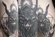
The tattoo of the goat, the photo of which are presented below, looks very eccentric and can be filled with a deep meaning. ABOUT...

Tattoo Art! How interesting it is interesting and multifaceted. Masterpieces created by masters conquer a huge ...

Abyssinian Cat (ABYSSINIAN CAT) Slender, medium-sized Abyssinian cat, gets well like ...
Sketches of small tattoos, in theory, the ideal task for the wizard! Draw - not much, which means, quickly and ...

Fatty painting today is very popular. But not everyone likes to apply huge knockers on the back, neck or ...

- Here you say that a person cannot in itself understand that it is good that it's bad that the whole thing is in the environment that ...

Many people choose colors tattoo only because they look great on the body. However, for those who ...

Piercing is a fashionable phenomenon among young people, which is piercing certain sections of a person or ...

Hello everybody! While new travels are just being prepared, I decided to remember the past and write about the trip in the Caucasus ...

An excellent indicator of masculinity and strength will be stylish male tattoos. They will be an excellent choice for ...

To see the presentation with pictures, design and slides, download its file and open in PowerPoint ...

How to prepare for the GTU? In order to answer this question, let's remember what GTO is ...